Fichier:EM of influenza virus.jpg

De Wikonsult
Sauter à la navigation Sauter à la recherche
Fichier d’origine(700 × 743 pixels, taille du fichier : 82 Kio, type MIME : image/jpeg)

Ce fichier provient de Wikimedia Commons. Il peut être utilisé par d'autres projets. Sa description sur sa page de description est affichée ci-dessous.

Description

Description
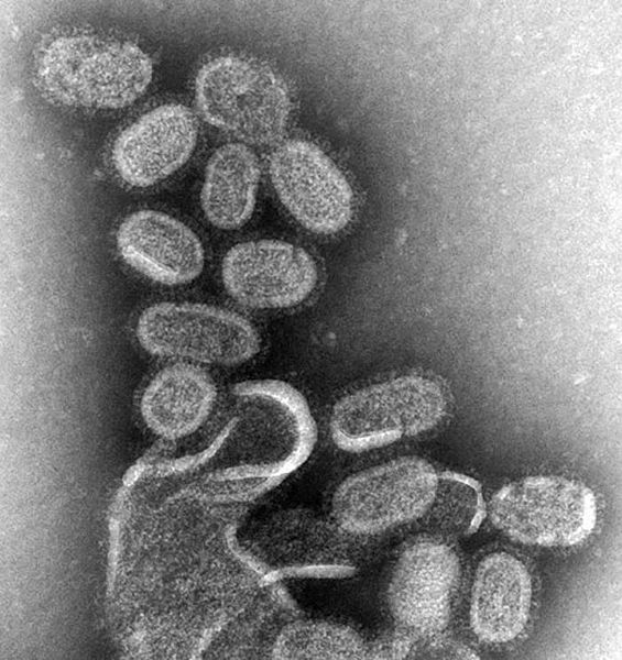
English: This negative stained transmission electron micrograph (TEM) shows recreated 1918 influenza virions that were collected from supernatants of 1918-infected Madin-Darby Canine Kidney (MDCK) cells cultures 18 hours after infection.

To separate these virions, the MDCK cells are spun down (centrifugation), and the 1918 virus in the fluid is immediately fixed for negative staining. The solid mass in lower center contains MDCK cell debris that did not spin down during the procedure.

Dr. Terrence Tumpey, one of the organization’s staff microbiologists and a member of the National Center for Infectious Diseases (NCID), recreated the 1918 influenza virus in order to identify the characteristics that made this organism such a deadly pathogen. Research efforts such as this, enables researchers to develop new vaccines and treatments for future pandemic influenza viruses.

The 1918 Spanish flu epidemic was caused by an influenza A (H1N1) virus, killing more than 500,000 people in the United States, and up to 50 million worldwide. The possible source was a newly emerged virus from a swine or an avian host of a mutated H1N1 virus. Many people died within the first few days after infection, and others died of complications later. Nearly half of those who died were young, healthy adults. Influenza A (H1N1) viruses still circulate today after being introduced again into the human population in the 1970s.
Ελληνικά: EM of influenza virus.jpg.
Tiếng Việt: siêu vi cúm qua hiển vi điện tử.
Date
Source

Ce document provient de la Bibliothèque d'images de santé publique (Public Health Image Library) (PHIL) des Centres pour le contrôle et la prévention des maladies , avec le numéro d'identification #8160.

Remarque : toutes les images de la PHIL ne sont pas dans le domaine public ; assurez-vous de vérifier le statut vis-à-vis des droits d'auteur et les crédits aux auteurs et aux fournisseurs de contenus.


العربية | Deutsch | English | македонски | slovenščina | +/−

Originally from en.wikipedia; description page is/was here.
Auteur
  • Photo Credit: Cynthia Goldsmith
  • Content Providers(s): CDC/ Dr. Terrence Tumpey
Autorisation
(Réutilisation de ce fichier)
PD-USGov-HHS-CDC
English: None - This image is in the public domain and thus free of any copyright restrictions. As a matter of courtesy we request that the content provider be credited and notified in any public or private usage of this image.

Conditions d’utilisation

Public domain
Cette image est l’œuvre des
Centers for Disease Control and Prevention
, division du Département de la Santé et des Services Sociaux des États-Unis, réalisée par un employé dans le cadre de ses activités professionnelles. En tant qu'œuvre du gouvernement fédéral des États-Unis d'Amérique, cette image est placée dans le domaine public.

Journal des téléversements d’origine

(All user names refer to en.wikipedia)

  • 2006-10-26 03:31 TimVickers 700×743×8 (83774 bytes) CDC, CDC Public Health Image Library (PHIL), http://phil.cdc.gov/Phil/details.asp

Légendes

Ajoutez en une ligne la description de ce que représente ce fichier
Influenza viruses under an electron microscope

image/jpeg

Historique du fichier

Cliquer sur une date et heure pour voir le fichier tel qu'il était à ce moment-là.

Date et heureVignetteDimensionsUtilisateurCommentaire
actuel10 août 2007 à 14:41Vignette pour la version du 10 août 2007 à 14:41700 × 743 (82 Kio)ToNToNi{{Information |Description=CDC, CDC Public Health Image Library (PHIL), http://phil.cdc.gov/Phil/details.asp |Source=Originally from [http://en.wikipedia.org en.wikipedia]; description page is/was [http://en.wikipedia.org/w/index.php?title=Image%3AEM_of_i

La page suivante utilise ce fichier :